Procedures
Arthroscopic Meniscectomy
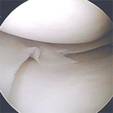
Arthroscopic meniscectomy is the most commonly performed of all arthroscopic operations. During surgery, a telescope is introduced into the knee which illuminates the knee joint cavity whilst filling it with fluid. This provides high quality images of the interior of the knee joint. The camera is usually introduced sequentially through three small wounds which are called arthroscopy portals, two made below the kneecap on either side of the patella tendon and a third above and outside the kneecap (this third wound is usually used to inspect the kneecap joint). The shock-absorbing pads of gristle, called the menisci, cup the knuckles of the joint supporting them as they slide and rotate upon each other. Tears of these meniscal cartilages are colloquially known as “torn cartilage”.
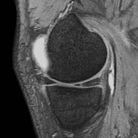
A severe injury can tear a cartilage from its root or bed. These types of injuries are found most often in children, adolescents and young adults, and sometimes they can be repaired with fine stitches. More usually, cartilages fail through areas of weakness that have developed within. This type of cartilage tear is most common in people in middle age. Symptoms can develop gradually or insidiously over a long period of time, occasionally they are the result of a twisting or stumbling injury. The presence of a meniscal tear is usually confirmed by imaging and often an MRI scan will be required. Arthroscopic meniscectomy normally involves removal of the torn piece of cartilage only, sparing as much normal cartilage as possible and contouring the cut edge to ensure that the knee runs smoothly following surgery.
Arthroscopy is not a particularly painful procedure, all patients are given painkillers to take home and most patients use these painkillers on the first day or two following surgery. Thereafter, they are not usually required.
Further information regarding the procedure and the aftercare can be found here.


